📊🔎 งานวิจัยใหม่บอกอะไรกับเรา
โรคหลอดเลือดสมอง หรือที่หลายคนเรียกว่า “สโตรก (Stroke)” ไม่ได้จบแค่ช่วงเวลาฉุกเฉิน แต่ยังอาจส่งผลระยะยาวต่อสมองมากกว่าที่คิด
งานวิจัยขนาดใหญ่จากสหรัฐอเมริกา ซึ่งติดตามผู้เข้าร่วมกว่า 42,000 คน เป็นเวลานานหลายปี พบว่า
👉 “ความรุนแรงของ Stroke” มีความสัมพันธ์โดยตรงกับ
- การเสื่อมของสมอง
- และความเสี่ยงในการเกิดภาวะสมองเสื่อม (Dementia)
📈🧠 ยิ่งรุนแรง สมองยิ่งเสื่อมเร็ว
ผลการศึกษาพบแนวโน้มที่ชัดเจนว่า
- 🟢 คนที่ไม่เคยเป็น Stroke → สมองเสื่อมตามวัยปกติ
- 🟡 Stroke ระดับเล็ก → สมองเริ่มเสื่อมเร็วขึ้น
- 🟠 Stroke ระดับปานกลาง → เสื่อมเร็วขึ้นชัดเจน
- 🔴 Stroke ระดับรุนแรง → สมองเสื่อมเร็วที่สุด
นักวิจัยเรียกสิ่งนี้ว่า “ความสัมพันธ์แบบเพิ่มตามระดับ (dose-response)”
หมายความว่า ยิ่งอาการรุนแรงมาก → ผลกระทบต่อสมองยิ่งมากตามไปด้วย
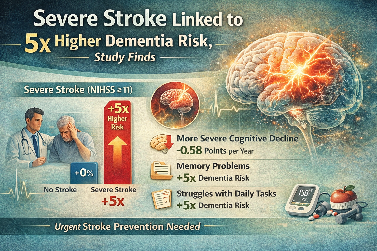
🚨📊 ความเสี่ยง “สมองเสื่อม” เพิ่มขึ้นหลายเท่า
เมื่อเทียบกับคนที่ไม่เคยเป็น Stroke
- 🔸 Stroke เล็ก → เสี่ยงสมองเสื่อมเพิ่มเกือบ 2 เท่า
- 🔸 Stroke ปานกลาง → เสี่ยงเพิ่มมากกว่า 3 เท่า
- 🔸 Stroke รุนแรง → เสี่ยงเพิ่มสูงถึง 5 เท่า
ตัวเลขนี้สะท้อนชัดว่า Stroke ไม่ใช่แค่ “อาการเฉียบพลัน”
แต่เป็นจุดเริ่มต้นของปัญหาสมองในระยะยาว
🧬💡 ทำไม Stroke ถึงกระทบสมองระยะยาว
นักวิจัยอธิบายแบบเข้าใจง่ายว่า
- 🧠 Stroke ทำให้ เนื้อสมองบางส่วนเสียหาย
- 🔗 ส่งผลต่อ “การเชื่อมต่อของสมอง” ที่ใช้คิด จำ และวางแผน
- 🔁 เพิ่มโอกาสเกิด Stroke ซ้ำ หรือโรคหลอดเลือดสมองขนาดเล็ก
- 🧍♂️ ผู้ป่วยอาจเคลื่อนไหวได้น้อยลง ทำให้ “การกระตุ้นสมองลดลง”
ทั้งหมดนี้ทำให้สมอง “ฟื้นตัวได้ยาก” และเสื่อมเร็วขึ้น
🧠📉 ไม่ใช่แค่ความจำ แต่กระทบหลายด้าน
ผลกระทบจาก Stroke ไม่ได้เกิดแค่เรื่อง “ลืมง่าย” เท่านั้น
แต่ยังรวมถึง
- ⚙️ การคิดวิเคราะห์และวางแผน
- 🧩 การตัดสินใจ
- 📊 การประมวลผลข้อมูล
ซึ่งเป็นทักษะสำคัญในชีวิตประจำวัน
🏥🛡️ สิ่งที่ควรรู้: ป้องกัน Stroke = ลดเสี่ยงสมองเสื่อม
ข้อค้นพบสำคัญของงานวิจัยนี้คือ
👉 การป้องกัน Stroke โดยเฉพาะ “Stroke รุนแรง”
สามารถช่วยลดความเสี่ยงสมองเสื่อมในอนาคตได้
วิธีดูแลตัวเองที่สำคัญ เช่น
- 🩺 ควบคุมความดันโลหิต
- 🍬 ควบคุมระดับน้ำตาลในเลือด
- 🚶♂️ ออกกำลังกายสม่ำเสมอ
- 🥗 กินอาหารที่ดีต่อสุขภาพ
- 🧠 สังเกตอาการความจำหลัง Stroke
🧭📌 สรุปแบบเข้าใจง่าย
- Stroke ยิ่งรุนแรง → สมองยิ่งเสื่อมเร็ว
- ความเสี่ยงสมองเสื่อมอาจเพิ่มสูงถึง 5 เท่า
- ผลกระทบเกิดได้หลายด้าน ไม่ใช่แค่ความจำ
- การป้องกันและดูแลสุขภาพ คือกุญแจสำคัญที่สุด
👉 กล่าวง่ายๆ คือ “ป้องกัน Stroke ได้ = ลดโอกาสสมองเสื่อมในอนาคต”
📚 แหล่งที่มา
- Koton S et al. Ischemic Stroke Incidence and Severity and Poststroke Cognitive Decline and Incident Dementia
- วารสาร JAMA Network Open (Published: April 16, 2026)
- DOI: 10.1001/jamanetworkopen.2026.8900
📄 เงื่อนไขการใช้งาน
บทความนี้เรียบเรียงจากงานวิจัยต้นฉบับ และเผยแพร่ภายใต้เงื่อนไข Creative Commons Attribution (CC-BY)
สามารถนำไปใช้ อ้างอิง หรือเผยแพร่ต่อได้ โดยต้องให้เครดิตแหล่งที่มาอย่างเหมาะสม.
📄 Terms of Use
This article is adapted from original research and is published under the Creative Commons Attribution (CC-BY) license.
It may be used, cited, or redistributed, provided that appropriate credit is given to the original source.